This article presents a fresh perspective on osteoarthritis, challenging the traditional view that the disease primarily originates in the bone. Instead, it argues that osteoarthritis begins with the tendons. The process starts with inflammation and shortening of the tendons, which gradually forces the joints closer together. Over time, this leads to bony hypertrophy and the development of osteoarthritis. Crucially, this mechanism is not confined to a single joint but can affect multiple areas of the body, reshaping our understanding of the disease and its progression.
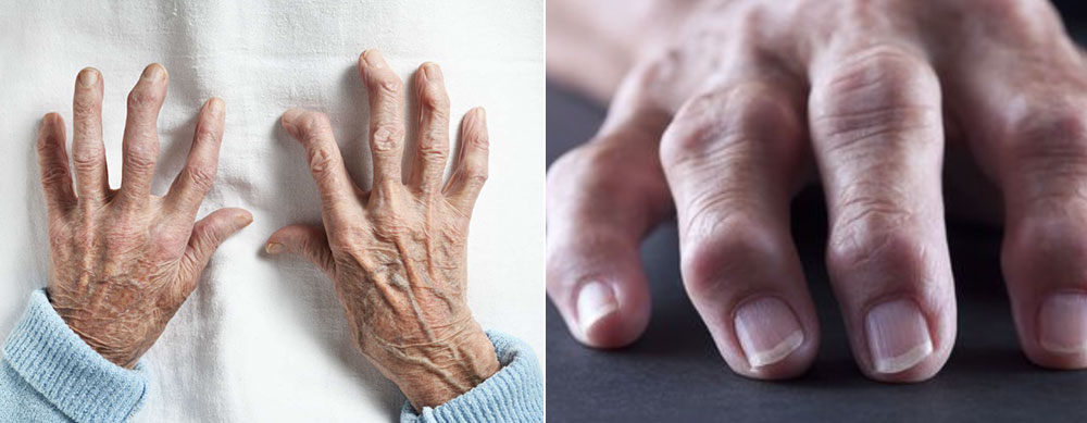
Clinicians, particularly Rheumatologists, are well-acquainted with Heberden’s and Bouchard’s nodes—bony hypertrophies or spurs at the level of the DIPs and PIPs of the fingers, which are hallmark signs of osteoarthritis. These changes are typically associated with the wear and tear of joints, affecting not only the fingers but also central, peripheral, and weight-bearing joints. Traditionally, osteoarthritis has been attributed to joint overuse and microtrauma, especially in the case of these nodules. However, the fact that these bony changes can also appear in the non-dominant hand suggests that osteoarthritis is not solely a result of repeated trauma or injury.
As a Rheumatologist with over 30 years of experience, I have observed that osteoarthritis can manifest even in individuals who rarely use their hands for strenuous tasks. Interestingly, osteoarthritis of the fingers often develops earlier than in weight-bearing joints, and there appears to be a familial tendency, with mother-daughter pairs sometimes exhibiting similar finger deformities. These deformities are typically characterized by the development of bony nodules, bone growth, and narrowing of the joint spaces. However, one notable aspect of finger deformity in osteoarthritis is the flexion or rotation of the last digit, which can occur even without the formation of nodules or bony hypertrophy.

While deformities are most noticeable at the last digit of the fingers, they are less apparent at the PIP joints and absent at the MCP level. In more advanced cases, the affected fingers may become partially or fully flexed at the PIP joint, with one or two fingers being more severely affected. This pattern suggests that osteoarthritis of the fingers is not solely a result of mechanical overuse, but may also be driven by deeper, systemic factors.
The development of bony hypertrophy is not simply a result of aging or injury; it is primarily caused by the shortening of the tendons, which leads to finger deformity and, eventually, osteoarthritis and bony hypertrophy at the DIPs and PIPs.
So, what exactly causes bony hypertrophy?
In a healthy, young hand, the flexor digitorum tendons are neither palpable nor tender. However, in certain individuals, these tendons can become inflamed, tender, and progressively fibrotic, eventually shortening. As the tendons shorten, they pull the fingers in an abnormal direction, leading to deformity. While inflammatory arthritis, such as rheumatoid arthritis (RA) or psoriatic arthritis (PsA), often presents with elevated inflammatory markers, pure tenosynovitis does not show these markers in laboratory studies. This distinction highlights that tendon shortening, rather than joint inflammation, is the primary driver of the observed changes in osteoarthritis.
When the tendon sheaths become thickened and swollen, they often lead to conditions like carpal tunnel syndrome. Tenosynovitis of the flexor digitorum tendons (FDT) differs from Dupuytren’s contracture, which typically affects only one or two fingers. Dupuytren’s contracture causes severe fibrosis that extends to the level of the MCP joints. Unfortunately, there is no cure for tenosynovitis. However, local application of NSAIDs, along with oral NSAIDs or intermittent steroid doses, can help manage symptoms.
While carpal tunnel release surgery can alleviate the paresthesia and numbness associated with carpal tunnel syndrome, it does not address the underlying tenosynovitis. Surgical release of the flexor tendons does not restore normal function or correct the deformities at the DIPs and PIPs, which have been caused by tendon shortening. In fact, surgery may exacerbate the problem, leading to further thickening and scarring of the flexor tendons.
The tendons and their sheaths, particularly in peripheral and weight-bearing joints, play a critical role in joint function. When these structures become inflamed and scarred, they shorten, progressively drawing the joints closer together, which can result in bone damage and the development of osteoarthritis.
Tenosynovitis can ultimately lead to the shortening of tendons, which may result in tendon ruptures, such as the rotator cuff, as seen in the image below. When the rotator cuff becomes inflamed and fibrotic, it can eventually rupture, leading to significant joint dysfunction.
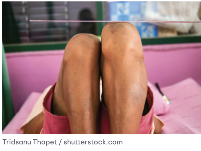
Similarly, leg length discrepancies can cause uneven pressure on the joints, contributing to unilateral osteoarthritis in the knee. This discrepancy, along with other factors like microtrauma, excess weight, and joint deformities, all increase the likelihood of developing osteoarthritis.
Upon examining the peripheral joints, one can observe the significant role tendon sheaths play in joint function. These sheaths, when inflamed, gradually shorten, ultimately contributing to the development of osteoarthritis.
In older individuals, height loss is commonly attributed to osteoporosis, osteoarthritis, and compression fractures. However, it’s important to recognize that shortening of the spine, as well as the inflammation and shortening of the paravertebral tendon sheaths, also contribute significantly to spinal height loss.
This broader understanding of tendon inflammation and shortening ties back to the central idea of this article—that osteoarthritis is not merely a disease of the bones, but a progressive condition driven by tendon and soft tissue changes. From the rotator cuff to the spine, tendon dysfunction plays a pivotal role in the development of osteoarthritis, reshaping how we view and approach joint health.